Introduction
“The process by which the cells are grown outside the body of organism, in artificial environment as in laboratory is termed as cell culturing and the cells are termed as cell culture.”
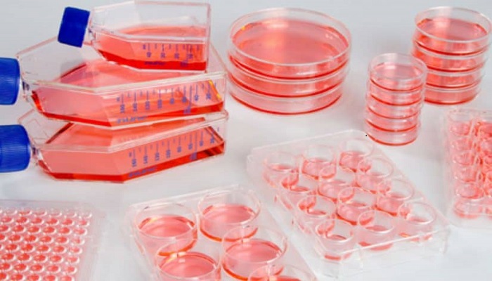
In laboratory for experimental and research purpose, the researcher grow cells under certain specific conditions. These are temperature, pH, concentration of certain gases as CO2 and O2 etc. Moreover, this require a specialized vessel that support the cell culture by providing them all the required nutrients for the growth. The cell culture now days are mainly a term use for the animal of the eukaryotic cell culture.
Steps Involved In Cell Culture Technique
The cell culture technique consists of these steps:
1. Isolation of cells
This involves the isolation of cells from animal tissues. The cells can be obtained from blood as well as isolated from the tissue digestion. Based upon the type of cells being cultured, this method may be different. For example, for cell culture of white blood cells the cells obtained from blood are more useful.
2. Maintaining the cell culture
The cells are grown in a specialized vessel that contains all essential nutrients for cell growth, optimum temperature and gas concentrations. The conditions and the growth medium may vary. Most widely used culture media are fetal bovine serum (FBS), Bovine calf serum and serum obtained from blood etc. Human platelet lysate (HPL) is another better option for cell culture media.
So these are the basic steps of the cell culturing technique. The development of this cell culturing technique is based upon several events and trial of scientists to culture the eukaryotic cells in vitro. So the history of cell culturing technique is as follow:
History of Animal Cell Culture
Year 1878
Scientist Claude Bernard proposed the possibility of maintaining physiological systems of the organism even after the death of organism, in a living system.
Year 1885
Roux maintained the embryonic chick cells in saline culture.
Year 1897
The survival of cells isolated from blood and connective tissue in plasma and serum was demonstrated by Loeb.
Year 1903
Observation and detailed study of in vitro cell division of salamander leucocytes.
Year 1907
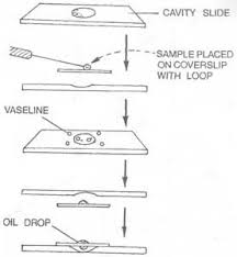
Ross Harrison cultivated the nerve cells of frog by the hanging drop method. On these bases, some consider him as the Father of cell culture technique.
The hanging drop method is a type of wet-mount technique in which the cells of organism to be cultured are suspended in a liquid droplet. Later, this droplet is placed on microscopic slide to observe their motility and cell division.The cover slip of microscope contains some sticky substance like petroleum jelly that prevents evaporation of liquid material.
Year 1910
Barrows an his team extended the work of Harrison. He cultivated the chicken embryo cells using cells of plasma clots and studied the process of mitosis in cells.
Year 1911
The very first liquid media for supporting the cell culture was prepared by Lewis using sea water, salts, peptones, serum and embryo extract. This liquid media provided monolayer growth of cells.
Year 1912
Alexis Carrel performed the cell culture technique in which the cell proliferation was enhanced by using liquid media, consisting of tissue and chick embryo extract. The liquid culture media provided all essential growth nutrients and fibrin clot of plasma provided the solid attachment surface for cell for better maintenance of cell culture. The use of trypsin for degradation or isolation of cells from the animal tissue was vital in this technique.
Year 1913
One major issue during cell culture technique was contamination of cell culture from the serum and liquid media. These basically serve as the support media. The discovery of aseptic techniques and antibiotic improved the cell culture technique.
Carrel introduced strict aseptic techniques for maintaining tissue culture for longer time period. He maintained the cells of chick embryo’s heart and it continued beating for three months. This was quite a longer time period just because of his aseptic techniques to prevent contamination of cell culture.
Human carcinoma cells (HeLa cells) were isolated and grown in vitro.
Year 1916
The proteolytic enzyme trypsin was introduced by Rous and Jones. Typsin enzyme is useful as it helps in dissociation of cells from the adherent surface of vessel in which they are cultured. This technique is termed as trypsinization as this enzyme digests the proteins that are responsible for adherence of cells to the culture medium
Year 1923
Specialized vessel was developed by Bakers and Carrel to culture animal/ eukaryotic cells and this vessel was useful for microscopic analysis of cell culture.
The cell culture vessel consists of adherent surface for the cells that need some support surface to proliferate and a specialized media that contains all essential nutrients required for cell growth. The media is highly specific depending upon the type of culture. For example, the Dulbecco modified Eagle medium (DMEM) etc.
The vessel provided support surface and essential nutrient media to cells. The tilted opening of vessel prevents leakage of media and prevents contamination.
Year 1927
Researchers Rivera and Carrel developed the first viral vaccine, Vaccinia.
Year 1933
Development of roller tube technique by Gay.
In fact, the researchers developed this technique for proper aeration of the cell culture in tube. The culture tubes were held in the rolling machine that would give them a turn in one hour for aeration.
Year 1940
Poliomyelitis virus was cultured for the first time by Weller, Robbins and Enders. The HeLa cell culture was useful as it provided a way to test certain chemicals and to observe the multiplication of viral particles in living cells of host.
Late in 1940’s the use of antibiotics, streptomycin and penicillin and the other aseptic techniques to prevent the contamination of cell culture was a great step in cell culturing technique.
Year 1948
Earle isolated and cloned the cells of mouse L fibroblasts. Moreover, Fischer prepared and introduced a chemically defined medium, CMRL 1066.
Year 1949
Enders proposed that the growth of Polio virus is possible using human embryonic cells as growth media.
So the preparation of polio vaccine and the syntactic medium for cell culture were events in animal cell culture technology. This synthetic media was either serum or fetal calf cells that consisted of all essential nutrients vitamins and other supplements to support cell culture.
Year 1952
Gey developed a continuous cell line of the human cervical carcinoma cells also termed as Hela cells or HeLa lane.
Researchers developed a plaque assay of animal viruses on monolayer of cultured cells.
Year 1954
Abercrombie observed the phenomenon of contact inhibition; that the motility of the diploid cells ceases when they come in contact to adjacent cells.
Year 1955
Eagle observed the nutritional requirements of the cell culture and also developed the first chemically defined medium.
Year 1961
Researchers Hay Flick and Moorhead isolated Human fibroblasts cells line (WI-38). They proposed that these cells have finite life-span.
Year 1964
Littlefield introduced the HAT medium for selection of cells.
Year 1965
Development of first serum- free medium.
The same year, Harris and Watkins experimented on fusion of human and the mouse cells, using viruses.
Year 1975
Milstein and Kohlar developed the very first hybridoma. This cell line developed and secreted monoclonal antibodies.
The Hybridoma Technique creates identical, cloned antibodies. Therefor, these antibodies are also termed as monoclonal antibodies. The organism’s body produce the in response to some antigen injected in them.
Year 1978
The researchers established the basis for development of serum free media, using hormones and some other growth factors.
Year 1982
Development of Human insulin, the first recombinant protein as therapeutic agent.
Year 1985
Human growth hormones were obtained from the recombinant bacteria. These are useful as therapeutic agent.
Year 1986
Licensing of Lymphoblastoidy YIFN.
These Lymphoblastoidy YIFN cell lines are produced by infection of human B-cells by EBV virus. The result is immortalization of cells and these are a source of producing transformed viral particles.
Year 1987
Development of Plasminogen activator for (TPA) from recombinant cell at commercial level.
Year 1989
Recombinant erythropoietin was still in trial.
Year 1990
Certain recombinant products were in clinical trials. For example, CD4, Factor VIII, HIVgp120 etc. The years after 1990 are also important as the experiments for the animal cloning were going on.
Year 1997
In 1997, the cloning experiment for animals was successful and scientists were able to clone a transgenic sheep, dolly by nuclear transfer technique.
The research is still going on for cloning of certain genes and the whole organism. But there are certain ethical issues as well. The national institute of immunology and the center for cellular and molecular biology are from those institute that are trying their best in cloning technique to develop certain type of cells so that the mutated cells in human body can be replaced by the normal cells.
Year 2006
Shinya Yamanaka and his colleagues demonstrated the phenomenon of induced pluripotent stem cells (iPS). The demonstrated the possibility of reprogramming of adult mouse fibroblast cells to wards the stem cell by simultaneous induced expression of four transcription factors. These include transcription factors Oct3/4, Sox2, C-Myc and Klf4. A very important trait of human iPS was their capability to form three germ layers in tissue culture an transplantation. However, there were morphological differences between natural stem stem cells of human body and that of iPS.
Year 2009
Until the year 2009, the researchers described the human ips resemble to embryonic stem cell (ES). They shared close morphology, expression of cell surface receptors, proliferation and genetic expression as well chromatic organization.
In 2009, Chin and colleagues presented results of their detailed study on both human iPS cell lines and embryonic stem cells. The results revealed differences in hundreds of gene expression.
Year 2012
John B. Guordon awarded with the Nobel Prize in physiology and medicine for their work that proved reversible nature of cell specialization.
These discoveries are great as they may help in future oncology and regenerative medicine. These also contribute in hematopoietic disease therapies.
Year 2012 to onwards
The coming years were of advancements in cell culture technology. One of them is 3D bioprinting technique.
3D Bioprinting techniology
While studying and experimenting the cell-cell interactions or effects of medicines or therapies on cell in vitro, the researcher have problems in mimicking the in vivo state of cell. The monolayer of cells usually cannot maintain the architecture and pattern of in vivo state.
Therefore, this 3D bioprinting developoment helped to create cell culture microenvironments similar to in vivo. This is useful and applicable both for adherent and non adherent cell cultures. As it maintains the cell- cell interaction, their architecture, proliferation and morphological features, this technique is useful for oncological studies and cell culture studies. Moreover, this will help in much accurate results and scafflod free 3D cell culture models for
Bottom line
The cell culture technique of genetic engineering has wide application in field of medicine and therapeutics. For instance, this will pave ways for better vaccine technology, recombinant protein therapeutics and gene therapy for treatment of genetic disorders. The stem cell technology has significance to replace the traditional treatment therapies for cancer treatment and inherited genetic disorders.