
Radioimmunoassay is an assay technique for detection and estimation of immune molecule complexes, antibodies, hormones and related substances from a given sample. This is sensitive and specific in vitro technique for research work laboratories. Because of the fact that this technique involves using radioactive isotopes, one needs great expertise to use this technique. Another example of radioimmunoassay is radioallergosorbent (RAST). This technique is specific for detection and estimation of allergens in blood.
Rosalyn Sussman Yalow and colleagues developed this technique at Veteran Administration Hospital, Bronx, New York. Dr. Yalow was awarded with noble prize in medicine in year 1977 for this technique.
This technique is based on the principle that radioactive labeled antigen molecules bind to particular antibodies due to their high affinity for each other. The level of radioactivity of supernatant provides assay of specific antibodies.
Radioimmunoassay (RIA) is an in-vitro technique of measuring or assaying an antigen in given solution with very high sensitivity for that antigen. In fact, almost all the biological substances, enzymes and proteins can be measured with this detection assay or technique.
Steps in radioimmunoassay (RIA)
In radioimmunoassay (RIA) technique, the target antigen is radioactively labeled and due to its affinity, it is able to bind to specific antibodies. Of course, a limited or known amount of specific antibodies is added. Adding a sample, such as blood-serum is added to initiate the competitive reaction. This competition is in between the labeled and unlabeled antigens to bind to the specific antibodies (whose detection is required).
As the reaction proceeds, both the labeled and unlabeled antigens bind to antibodies molecules. Because of this competition, some of these will remain unbound and will be present in free form in solution. The amount of both types of antigens is estimated. A curve is plotted of the ratio of both antigens.
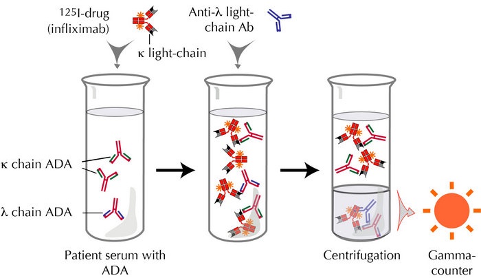
In next step, the bound antigens are separated from the unbound. The radioactivity of free antigens is measured from the supernatant. This radioactivity is used for assaying the detection and amount of desired protein or antibody molecules.
Requirements
While performing this technique, some requirements are:
- Specific antiserum to antigens for estimation
- Radioactively labeled antigens
- Some technique to separate the bound and unbound antigen-antibody complexes
- Instrumentation to measure and estimate radioactivity level
Source of radioactivity
In order to create a radio-labeled antigen, certain radioactive isotopes are used. For instance, C14, H3 and I-125 antigens. The radiolabel 125-I is frequently used for radioimmunoassay. This highly specific and efficient radio-labeled antigen is prepared by iodination of pure antigen. The radioactive isotope usually binds at tyrosine residue. Either Chloramines-T or peroxidase method is used for making this labeling possible. Later on, high performance liquid chromatography (HCL) separates the labeled antigens
Separation of bound and unbound antigens
Certain separation technique is used to separate the radio-labeled antigens to be used in radioimmunoassay later. These techniques are:
- Double antibody
- Cellulosic membrane
- Chromatography
- Solid- phase technique
Amongst these, double antibody technique in combination with polyethylene is most frequently used. The gamma counter counts and measures the bound antigen. The final step is plotting a standard curve or calibration graph to calculate the amount of unknown (antibody or protein product) entity.
Disequilibrium technique improves the sensitivity of these radio-labeled antigens. This technique applies a short incubation period for antigen and antibody molecules. While the radioactivity is applied later. Radioimmunoassay is relatively older technique, yet still in use for many laboratory procedures as detection and assay technique.
Applications
This technique has wide applications such as:
- Detection of drugs such as narcotics and morphine in hair samples
- Estimation of mere amounts of hydromorphone and hydrocodone derivatives of morphine drug in blood sample
- Detecting Flunisolide in blood plasma. Flunisolides is corticoid and designed for treatment of asthma, allergy and arthritis.
- Estimation of Ferritin in blood serum of patient. This Ferritin in high concentration is indicative of iron stores. Sometimes the body has the sufficient levels of iron yet the stored form of iron is not used for some metabolic condition. Therefore, RIA in such case is helpful to identify if there is truly iron deficiency in patient body or some metabolic issues.
- Thyroids testing by estimating the levels of thyroxin in blood
- Detection of certain toxins in blood sample
your blog is very knowlageble.